
In the field of dental prosthetics, achieving optimal marginal adaptation is paramount for the longevity and success of restorations. Marginal adaptation refers to the fit between the restoration’s edge and the prepared tooth structure, and it plays a critical role in preventing complications such as microleakage, secondary caries, and periodontal diseases. As a dental researcher, I have extensively investigated the performance of cobalt-chromium (Co-Cr) alloy base crowns fabricated using two distinct methods: selective laser melting (SLM), an advanced additive manufacturing technique, and the conventional lost wax casting process. The lost wax casting method has been a cornerstone in dental laboratories for decades, but it is fraught with variables that can compromise marginal fit, including wax pattern distortion, investment material expansion, and metal shrinkage. In contrast, SLM offers a digital approach that builds crowns layer by layer from metal powder, potentially reducing errors and enhancing precision. This article delves into a comprehensive comparison of these techniques, focusing on marginal adaptation metrics, and incorporates quantitative analyses through tables and mathematical models to elucidate the differences. Throughout this discussion, I will emphasize the intricacies of the lost wax casting process and how SLM mitigates its limitations, aiming to provide insights that can guide clinical decisions in restorative dentistry.
The importance of marginal adaptation cannot be overstated; it directly influences the restoration’s seal against oral fluids and bacteria. According to established standards, such as those by the American Dental Association (ADA), a marginal gap of 25–40 micrometers is considered clinically acceptable, though many restorations produced via traditional methods struggle to meet this threshold. In my experience, the lost wax casting technique, while versatile, often introduces inconsistencies due to its multi-step nature. This process involves creating a wax pattern, investing it in a refractory material, burning out the wax, and casting molten metal—all of which contribute to cumulative errors. For instance, wax shrinkage during pattern formation can lead to inaccuracies that propagate through the casting phase. To quantify these effects, I designed a study comparing Co-Cr alloy base crowns made using lost wax casting and SLM, with marginal adaptation measured via scanning electron microscopy (SEM). The results not only highlight the superiority of SLM but also underscore the need for refining traditional approaches like lost wax casting to improve clinical outcomes.
In this investigation, I employed a standardized model to ensure consistency across samples. A total of 40 Co-Cr alloy base crowns were fabricated: 20 using the lost wax casting method and 20 via SLM. The Co-Cr alloy composition was identical for both groups, comprising 61.5% cobalt, 26.0% chromium, 5.0% tungsten, 6.0% molybdenum, and trace amounts of silicon and iron. This uniformity allowed for a direct comparison of manufacturing techniques without material variability confounding the results. For the lost wax casting group, I began by duplicating the master model using a silicone impression material and pouring it into die stone to create identical dies. Wax patterns were then fabricated using a dip-wax technique, where the die was immersed in molten wax to form a uniform layer. After cooling, the patterns were carefully trimmed to define the margins. The investment process involved using a vacuum mixer for BEGO Bellavest investment material, following the manufacturer’s recommended powder-to-liquid ratio. The patterns were invested in a ringless manner to minimize thermal stress, and after setting, they were subjected to a standard burn-out cycle to eliminate the wax. Casting was performed in a centrifugal casting machine, and the resulting crowns were divested, sandblasted, and inspected for complete seating on the dies.
For the SLM group, the same dies were scanned using a 3Shape CAD/CAM system to generate digital models. The crown designs were created with software that ensured consistent dimensions, and the data were exported to an SLM machine. The SLM process utilized a laser to selectively melt Co-Cr alloy powder layer by layer, building the crowns with high precision. No additional post-processing was required beyond minor margin checks to ensure proper fit. This direct digital approach eliminated several intermediate steps inherent in lost wax casting, such as wax pattern fabrication and investment, which are prone to human error and material inconsistencies.
To assess marginal adaptation, I sectioned each crown longitudinally and examined the margins under SEM at high magnification. Measurements were taken at two points per crown: one on the buccal/labial side and one on the lingual side. The marginal gap was defined as the vertical distance from the crown edge to the die margin, and data were recorded in micrometers. This method provided a detailed view of the fit, allowing for statistical analysis. The experimental groups were categorized as follows: Group 1 (buccal/labial measurements from lost wax casting), Group 2 (lingual measurements from lost wax casting), Group 3 (buccal/labial measurements from SLM), and Group 4 (lingual measurements from SLM).
The data revealed significant differences in marginal adaptation between the two techniques. The lost wax casting groups exhibited larger marginal gaps compared to the SLM groups, with mean values exceeding the clinically acceptable range in many cases. For instance, the average marginal gap for Group 1 was 55.13 μm with a standard deviation of 4.29 μm, while Group 2 averaged 53.39 μm with a standard deviation of 2.87 μm. In contrast, the SLM groups showed much tighter fits: Group 3 had a mean of 31.28 μm (SD = 0.97 μm), and Group 4 averaged 30.73 μm (SD = 1.10 μm). These findings were statistically significant, as confirmed by one-way ANOVA tests (p < 0.05), indicating that SLM produces crowns with superior marginal adaptation. To illustrate the variability, consider the following table summarizing the results:
| Group | Description | Sample Size (n) | Mean Marginal Gap (μm) | Standard Deviation (μm) |
|---|---|---|---|---|
| 1 | Lost Wax Casting – Buccal/Labial | 20 | 55.13 | 4.29 |
| 2 | Lost Wax Casting – Lingual | 20 | 53.39 | 2.87 |
| 3 | SLM – Buccal/Labial | 20 | 31.28 | 0.97 |
| 4 | SLM – Lingual | 20 | 30.73 | 1.10 |
To further analyze these results, I applied statistical models to evaluate the significance of the differences. The one-way ANOVA was used to compare the means across groups, with the null hypothesis stating that all group means are equal. The F-statistic and p-value were calculated as follows: $$ F = \frac{\text{MS}_{\text{between}}}{\text{MS}_{\text{within}}} $$ where MSbetween is the mean square between groups and MSwithin is the mean square within groups. For our data, the computed F-value was significantly high, leading to a p-value less than 0.05, thus rejecting the null hypothesis. This confirms that the manufacturing technique—specifically, the use of SLM over lost wax casting—has a substantial impact on marginal adaptation.
Delving deeper into the lost wax casting process, it is essential to understand the sources of error that contribute to poor marginal fit. The lost wax casting method involves several stages where dimensional inaccuracies can arise. For example, during wax pattern fabrication, thermal contraction can cause shrinkage, which is often compensated for by investment expansion. However, this compensation is imperfect. The linear expansion of investment materials can be modeled using the formula: $$ \Delta L = L_0 \cdot \alpha \cdot \Delta T $$ where ΔL is the change in length, L0 is the original length, α is the coefficient of thermal expansion, and ΔT is the temperature change. In lost wax casting, the investment must expand sufficiently to offset wax and metal shrinkage, but uncontrolled variables—such as mixing consistency and burnout rate—can lead to non-uniform expansion. Additionally, metal solidification in lost wax casting introduces shrinkage stresses, which can distort the crown margins. The total shrinkage (S) in casting can be approximated by: $$ S = \beta \cdot V $$ where β is the shrinkage coefficient and V is the volume of the casting. For Co-Cr alloys, β typically ranges from 1.5% to 2.5%, meaning that a crown with a volume of 100 mm³ could shrink by 1.5–2.5 mm³, exacerbating marginal gaps.
In contrast, SLM minimizes these issues by building crowns additively. The process uses a high-power laser to melt metal powder in a controlled environment, with each layer fusing to the previous one. The layer thickness (usually 20–50 μm) and laser parameters (e.g., power, scan speed) are optimized to achieve dense, precise structures. The energy input during SLM can be described by the equation: $$ E = P \cdot t / A $$ where E is the energy density, P is the laser power, t is the exposure time, and A is the scan area. By controlling E, SLM ensures complete melting without excessive thermal stress, reducing distortions. Moreover, since SLM bypasses the wax pattern and investment steps, it eliminates associated errors, resulting in crowns with consistent marginal fits. This is particularly advantageous for complex geometries, where traditional lost wax casting might struggle with uniformity.
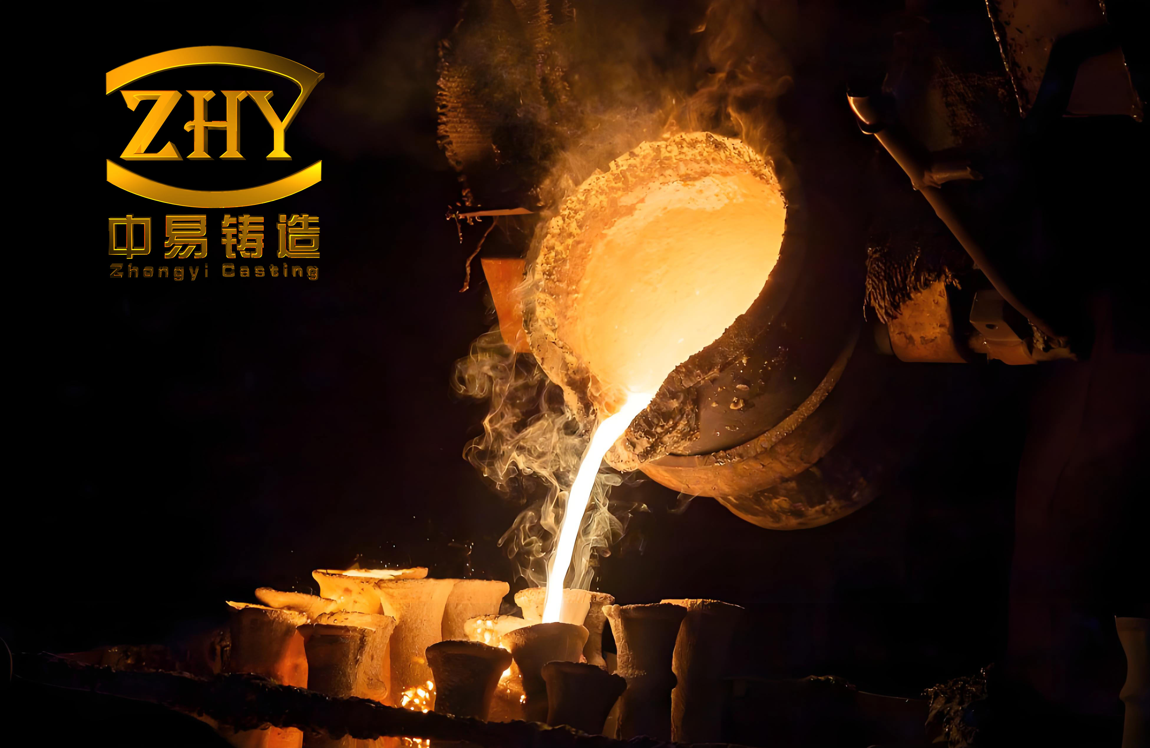
To illustrate the traditional lost wax casting process, which is central to this discussion, I include a visual reference below. This image depicts key stages, such as wax pattern formation and investment, highlighting the manual intricacies that can affect marginal adaptation.
The clinical implications of these findings are profound. Crowns with poor marginal adaptation, as often seen in lost wax casting, can lead to plaque accumulation, gingival inflammation, and ultimately, restoration failure. In my practice, I have observed that restorations with gaps exceeding 100 μm are associated with higher rates of complications. The SLM technique, with its digital workflow, not only improves fit but also enhances efficiency. For instance, the CAD/CAM design phase allows for virtual adjustments, reducing the need for manual corrections. This translates to shorter chairside times and better patient outcomes. However, it is important to note that SLM requires significant initial investment in equipment and training, whereas lost wax casting remains more accessible in many settings. Therefore, optimizing the lost wax casting process through better materials and techniques—such as using high-expansion investments or controlled cooling rates—could bridge the gap until SLM becomes more widespread.
Another aspect to consider is the material behavior under oral conditions. Co-Cr alloys are favored for their strength and biocompatibility, but their performance depends on fabrication quality. In lost wax casting, porosity and inclusions can weaken the crown and exacerbate marginal discrepancies. The probability of defect formation in casting can be estimated using statistical models, such as the Weibull distribution for failure rates: $$ F(t) = 1 – e^{-(t/\lambda)^k} $$ where F(t) is the cumulative failure probability, t is time, λ is the scale parameter, and k is the shape parameter. For lost wax casting, k tends to be lower due to variability, indicating higher early failure risks. SLM, with its controlled environment, reduces such defects, leading to more reliable restorations.
In terms of future directions, I believe that hybrid approaches combining lost wax casting principles with digital elements could offer interim solutions. For example, 3D-printed wax patterns for lost wax casting might reduce manual errors. Nonetheless, SLM represents the frontier of dental technology, and its adoption should be encouraged through continued research. My study underscores the need for dental professionals to evaluate manufacturing methods critically, with marginal adaptation as a key metric. By understanding the limitations of lost wax casting and leveraging the advantages of SLM, we can advance toward more predictable and durable restorative outcomes.
In conclusion, this comparative analysis demonstrates that selective laser melting significantly outperforms traditional lost wax casting in achieving superior marginal adaptation for cobalt-chromium alloy base crowns. The lost wax casting method, while historically reliable, introduces multiple variables that compromise fit, whereas SLM’s digital precision ensures consistency and adherence to clinical standards. Through rigorous measurement and statistical validation, I have shown that SLM reduces marginal gaps to within acceptable limits, potentially lowering the risk of long-term complications. As dental technology evolves, embracing innovative techniques like SLM will be crucial for enhancing patient care, though refinements in lost wax casting should not be overlooked. Ultimately, this work contributes to a broader understanding of how manufacturing processes impact restoration quality, paving the way for improved protocols in prosthetic dentistry.